Protective effects of Pithecellobium dulce seed extract on sodium fluoride-induced neurotoxicity in rats
DOI:
https://doi.org/10.69857/joapr.v14i4.2027Keywords:
Fluoride, Histology, Antioxidant, DPPH, Glutathione Peroxidase, NeurodegenerationAbstract
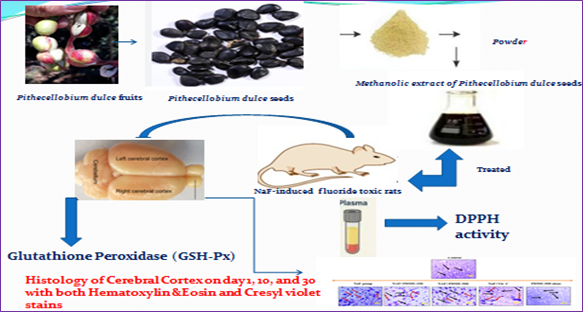
Background: According to epidemiological research, fluoride is a developmental neurotoxicant that lowers children's IQ scores. Fluoride induces oxidative stress in the brain, which can cause histological damage. This study aims to test the antioxidant potential of the methanolic extract of Pithecellobium dulce seeds. Methodology: By using different in vivo assays such as inhibition of DPPH activity in plasma, Glutathione Peroxidase (GSH-Px) activity in the brain, and histology of the cerebral cortex on days 1, 10, 20, and 30 with both Hematoxylin &Eosin and Cresyl violet stains. Results and Discussion: The methanolic extract of P dulce seeds significantly enhances GPx activity in a dose-dependent manner, effectively countering oxidative stress and providing neuroprotective benefits against fluoride-induced neurodegeneration. Results also showed the strong antioxidative potential of PDME, particularly in mitigating NaF-induced oxidative stress, as evidenced by sustained high DPPH radical-scavenging activity in the treated groups over time. An H&E histology study highlights the neuroprotective effects of PDME against NaF-induced neurodegeneration, demonstrating that PDME treatment significantly reduces neuronal loss and preserves cellular integrity, particularly at higher doses. Cresyl violet stain demonstrates that PDME has a neuroprotective effect against NaF-induced neurotoxicity, as evidenced by the preservation of neuronal morphology and Nissl substance. The study demonstrates that the methanolic extract of Pithecellobium dulce seeds exhibits significant antioxidant activity, effectively counteracting fluoride-induced neurotoxicity and preserving neuronal integrity by enhancing glutathione peroxidase activity and improving histological outcomes. Conclusion: These findings suggest that P. dulce may serve as a potential neuroprotective agent against fluoride-related cognitive impairments.
Downloads

References
Atanasov AG, Waltenberger B, Pferschy-Wenzig E, Linder T, Wawrosch C, Uhrin P, et al. Discovery and resupply of pharmacologically active plant-derived natural products: a review. Biotechnol Adv, 33, 1582–1614 (2015) https://doi.org/10.1016/j.biotechadv.2015.08.001
Shahidi F, Ambigaipalan P. Phenolics and polyphenolics in foods, beverages, and spices: antioxidant activity and health effects — a review. J Funct Foods, 18, 820–897 (2015) https://doi.org/10.1016/j.jff.2015.06.018
Liao BY, Zhu DY, Thakur K, Li L, Zhang JG, Wei ZJ. Thermal and antioxidant properties of polysaccharides sequentially extracted from mulberry leaves (Morus alba L.). Molecules, 22(12), 1–14 (2017) https://doi.org/10.3390/molecules22122271
Zhang YY, Zhang F, Thakur K, Ci AT, Wang H, Zhang JG. Effect of natural polyphenol on the oxidative stability of pecan oil. Food Chem Toxicol, 119, 489–495 (2018) https://doi.org/10.1016/j.fct.2017.10.001
Martins E. The growing use of herbal medicines: issues relating to adverse reactions and challenges in monitoring safety. Front Pharmacol, 4, 177–189 (2014) https://doi.org/10.3389/fphar.2013.00177
Nagmoti DM, Kothavade PS, Bulani VD, Gawali NB, Juvekar AR. Anti-diabetic and anti-hyperlipidemic activity of Pithecellobium dulce (Roxb.) Benth seeds extract in streptozotocin-induced diabetic rats. Eur J Integr Med, 7, 263–273 (2015) https://doi.org/10.1016/j.eujim.2015.01.001
Vargas AF, Kuri GA, Vargas MH, Chavez SJL, Ferriz RA, Hernandez SLG, et al. Phenolic profile and antioxidant capacity of Pithecellobium dulce (Roxb) Benth: a review. J Food Sci Technol, 57(12), 4316–4336 (2020) https://doi.org/10.1016/j.eujim.2015.01.001
Palika W, Piriya C, Chuchard P, Sineenart S. LC-QTOF-MS characterization, antioxidant activity, and in vitro toxicity of medicinal plants from the Tri-Than-Thip remedy. Evid Based Complement Alternat Med, 2022, 4477003 (2022) https://doi.org/10.1155/2022/4477003
Patil RP, Chaudhari RM, Pawer SP. A systemic review on Pithecellobium dulce (Jangal Jalaebi). Asian J Res Pharm Sci, 16(2), 167–171 (2026) https://doi.org/10.52711/2231-5659.2026.00026
Kumari S. Evaluation of phytochemical analysis and antioxidant and antifungal activity of Pithecellobium dulce leaves' extract. Asian J Pharm Clin Res, 10(1), 370 (2017) https://doi.org/10.22159/ajpcr.2017.v10i1.15576
Uma Shankar K, Prem Kumar A. Phytochemical screening, antimicrobial and anti-inflammatory activity of crude seeds extract from Pithecellobium dulce (Roxb.) Benth. Int J Pharma Bio Sci, 10(3), 26–30 (2019) https://doi.org/10.22376/ijpbs.2019.10.3.b26-30
Sara SK, Iriny MA, Safaa A, Moghazy EA, Nasser BS. Process optimization and characterization of Manila tamarind seed oil extracted by the Soxhlet method. Int J Energy Clean Environ, 22(1), 31–39 (2021) https://doi.org/10.1615/InterJEnerCleanEnv.2020034890
Rotruck JT, Pope AL, Ganther HE, Swanson AB, Hafeman DG, Hoekstra WG. Selenium: biochemical role as a component of glutathione peroxidase. Science, 179(4073), 588–590 (1973) https://doi.org/10.1126/science.179.4073.588
Pleban P, Munyani A, Beachum J. Determination of selenium concentration and glutathione peroxidase activity in plasma and erythrocytes. Clin Chem, 28, 311–316 (1982)
Polile RP, Matamane RP, Tlou L. Assessment of 2,2-diphenyl-1-picrylhydrazyl (DPPH) radical scavenging, total antioxidant activity, ferric reducing power and phytochemical analysis of methanolic extract of Malva parviflora. Journal of Natural Sciences Research, 15, 27-34 (2024) https://doi.org/10.7176/JNSR/15-2-03
Bradford MM. A rapid and sensitive method for the quantitation of microgram quantities of protein utilizing the principle of protein-dye binding. Anal Biochem, 72(1–2), 248–254 (1976) https://doi.org/10.1016/0003-2697(76)90527-3
Gornall AG, Bardawill CJ, David MM. Determination of serum proteins by means of the biuret reaction. J Biol Chem, 177(2), 751–766 (1949)
Mirzaei F, Khazaei M, Komaki A, Amiri I, Jalili C. Virgin coconut oil (VCO) by normalizing NLRP3 inflammasome showed potential neuroprotective effects in amyloid-β induced toxicity and high-fat diet fed rat. Food Chem Toxicol, 118, 68–83 (2018) https://doi.org/10.1016/j.fct.2018.04.064
Andersen JK. Oxidative stress in neurodegeneration: cause or consequence? Nat Med, 10(S7), S18–S25 (2004) https://doi.org/10.1038/nrn1434
Nahar L, Charoensup R, Kalieva K, Habibi E, Guo M, Wang D, et al. Natural products in neurodegenerative diseases: recent advances and future outlook. Front Pharmacol, 16, 1529194 (2025) https://doi.org/10.3389/fphar.2025.1529194
Mittal P, Goyal R, Kapoor R, Wan C, Gautam RK. Natural products-based drugs: potential drug targets against neurological degeneration. Curr Neuropharmacol, 21(4), 777–786 (2023) https://doi.org/10.2174/1570159X21666230220102605
Apetz N, Munch G, Govindaraghavan S, Gyengesi E. Natural compounds and plant extracts as therapeutics against chronic inflammation in Alzheimer's disease — a translational perspective. CNS Neurol Disord Drug Targets, 13(7), 1175–1191 (2014) https://doi.org/10.2174/1871527313666140917110635
Malatesta M. Histological and histochemical methods — theory and practice. Eur J Histochem, 60(1), 2639 (2016) https://doi.org/10.4081/ejh.2016.2639
Braak H, Braak E. Neuropathological stageing of Alzheimer-related changes. Acta Neuropathol, 82(4), 239–259 (1991) https://doi.org/10.1007/BF00308809
Schmued LC, Hopkins KJ. Fluoro-Jade B: a high affinity fluorescent marker for the localization of neuronal degeneration. Brain Res, 874(2), 123–130 (2000) https://doi.org/10.1016/s0006-8993(00)02513-0
Nakaso K, Kitayama M, Kimura K, Yanagawa T, Ohama E, Nakashima K, et al. Induction of heme oxygenase-1 in the rat brain by kainic acid-mediated excitotoxicity: the dissociation of mRNA and protein expression in hippocampus. Biochem Biophys Res Commun, 259(1), 91–96 (1999) https://doi.org/10.1006/bbrc.1999.0724
Flora SJ, Pachauri V. Chelation in metal intoxication. Int J Environ Res Public Health, 7(7), 2745–2788 (2010) https://doi.org/10.3390/ijerph7072745
Mattson MP. Neuroprotective signaling and the aging brain: take away my food and let me run. Brain Res, 886(1–2), 47–53 (2000) https://doi.org/10.1016/s0006-8993(00)02790-6
Babanna N, Reddy Karnati. 9-Hexadecenoic acid rich HPLC fraction of Pithecellobium dulce methanolic seed extract exhibits potential anti-inflammatory activity by inhibiting IL-8, IL-6, and PGE2: phytochemical characterization, in vitro and in vivo evaluation. J Res Pharm, 27(5), 1734–1750 (2023) https://doi.org/10.29228/jrp.458
Nabavi SM, Nabavi SF, Habtemariam S, Moghaddam AH, Latifi AM. Ameliorative effects of quercetin on sodium fluoride-induced oxidative stress in rat's kidney. Ren Fail, 34(7), 901–906 (2012) https://doi.org/10.3109/0886022X.2012.687347
Dhanisha SS, Drishya S, Guruvayoorappan C. Traditional knowledge to clinical trials: a review on nutritional and therapeutic potential of Pithecellobium dulce. J Basic Clin Physiol Pharmacol, 33(2), 133–142 (2021) https://doi.org/10.1515/jbcpp-2020-0166
Chouhan S, Flora SJS. Effects of fluoride on the tissue oxidative stress and apoptosis in rats: biochemical assays supported by IR spectroscopy data. Toxicology, 254(1–2), 61–67 (2008) https://doi.org/10.1016/j.tox.2008.09.008
Bains JS, Hall ED. Antioxidant therapies in traumatic brain and spinal cord injury. Biochim Biophys Acta Mol Basis Dis, 1822(5), 675–684 (2012) https://doi.org/10.1016/j.bbadis.2011.10.017
Sharma C, Suhalka P, Sukhwal P, Jaiswal N, Bhatnagar M. Curcumin attenuates neurotoxicity induced by fluoride: an in vivo evidence. Pharmacogn Mag, 10(37), 61–65 (2014) https://doi.org/10.4103/0973-1296.126663
Brigelius-Flohé R, Maiorino M. Glutathione peroxidases. Biochim Biophys Acta Gen Subj, 1830(5), 3289–3303 (2013) https://doi.org/10.1016/j.bbagen.2012.11.020
Dringen R. Metabolism and functions of glutathione in brain. Prog Neurobiol, 62(6), 649–671 (2000) https://doi.org/10.1016/s0301-0082(99)00060-x
Clemente-Suárez VJ, Martín-Rodríguez A, Beltrán-Velasco AI, Rubio-Zarapuz A, Martínez-Guardado I, Valcárcel-Martín R, et al. Functional and therapeutic roles of plant-derived antioxidants in type 2 diabetes mellitus: mechanisms, challenges, and considerations for special populations. Antioxidants (Basel), 14(6), 725 (2025) https://doi.org/10.3390/antiox14060725
Chaudhary S, Pinky, Parvez S. Neuroprotective effects of natural antioxidants against branched-chain fatty acid-induced oxidative stress in cerebral cortex and cerebellum regions of the rat brain. ACS Omega, 7(43), 38269–38276 (2022) https://doi.org/10.1021/acsomega.2c00163
Verma A, Rawat AKS. Phytochemical analysis, antioxidant potential, and cytotoxic activity of extracts of Quisqualis indica L. J Appl Pharm Res, 13(3), 247–258 (2025) https://doi.org/10.69857/joapr.v13i3.1215
Cuanalo-Contreras K, Moreno-Gonzalez I. Natural products as modulators of the proteostasis machinery: implications in neurodegenerative diseases. Int J Mol Sci, 20(19), 4666 (2019) https://doi.org/10.3390/ijms20194666
Ashok A, Andrabi SS, Mansoor S, Kuang Y, Kwon BK, Labhasetwar V. Antioxidant therapy in oxidative stress-induced neurodegenerative diseases: role of nanoparticle-based drug delivery systems in clinical translation. Antioxidants, 11, 408 (2022) https://doi.org/10.3390/antiox11020408
Padayatty SJ, Katz A, Wang Y, Eck P, Kwon O, Lee JH, et al. Vitamin C as an antioxidant: evaluation of its role in disease prevention. J Am Coll Nutr, 22(1), 18–35 (2003) https://doi.org/10.1080/07315724.2003.10719272
Merghany RM, El-Sawi SA, Naser AFA, Ezzat SM, Moustafa SFA, Meselhy MR. A comprehensive review of natural compounds and their structure-activity relationship in Parkinson's disease: exploring potential mechanisms. Naunyn Schmiedebergs Arch Pharmacol, 398(3), 2229–2258 (2025) https://doi.org/10.1007/s00210-024-03462-4
Dey D, Roy Mukherjee D, Shoeb A, Biswas P, Santra S. A systematic review of Alzheimer's disease: exploring genetic and environmental risk factors, biomarkers, and future pharmacotherapy for cognitive decline and neurodegeneration. J Appl Pharm Res, 13(3), 17–35 (2025) https://doi.org/10.69857/joapr.v13i3.929
Patel SS, Udayabanu M. Urtica dioica extract attenuates depressive like behavior and associative memory dysfunction in dexamethasone induced diabetic mice. Metab Brain Dis, 31(1), 1–12 (2016) https://doi.org/10.1007/s11011-014-9480-0
Bancroft JD, Gamble M. Theory and Practice of Histological Techniques. 6th ed. Churchill Livingstone, Elsevier, China (2008) https://doi.org/10.1016/C2015-0-00143-5
Howland RH. Vagus nerve stimulation. Curr Behav Neurosci Rep, 1, 64–73 (2014) https://doi.org/10.1007/s40473-014-0010-5
Sofroniew MV, Vinters HV. Astrocytes: biology and pathology. Acta Neuropathol, 119(1), 7–35 (2010) https://doi.org/10.1007/s00401-009-0619-8
Barnham KJ, Masters CL, Bush AI. Neurodegenerative diseases and oxidative stress. Nat Rev Drug Discov, 3, 205–214 (2004) https://doi.org/10.1038/nrd1330
Shukla M, Singh A, Ghosh P, Chatterjee S, Singh P. Systematic review on evidence based therapeutic potential of Pithecellobium dulce for health benefits. Toxicol Int, 31(2), 249–256 (2024) https://doi.org/10.18311/ti/2024/v31i2/36360
Published
How to Cite
Issue
Section
Copyright (c) 2026 Babbanna Yelugudari, Bhaskar Nagilla, Pratap Reddy Karnati

This work is licensed under a Creative Commons Attribution-NonCommercial 4.0 International License.